Active zones and postsynaptic clusters at the neuromuscular junction
The synaptic active zone is the site of vesicle docking and NT release. Here is a photo of a synaptic active zone from U Texas's transmission electron microscopy department:

These can look differently depending on the species and type of neuron. Generally they are recognizable by a high linear electron density in the EM image and close apposition between pre and postsynaptic membranes. Sometimes, increasing the EM magnification is necessary to see them.
The enzyme protein phosphatase 2A is necessary for the development of structurally normal active zones across the cleft from glutamate receptor clusters. Visquez et al expressed a nervous system-specific transgene that lacks the protein phosphatase 2 catalytic subunit in fruit flies. These fruit flies had less enzyme activity and a ~ 30% reduction in antibody staining to active zones at neuromuscular junctions.
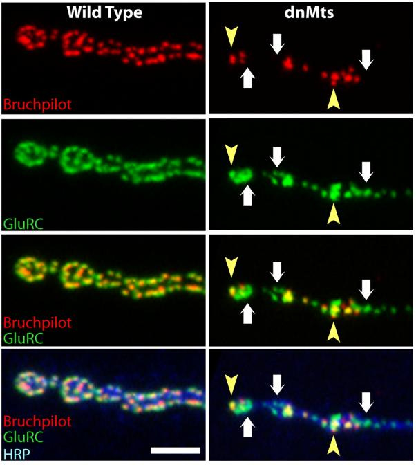
This system does not directly affect postsynaptic glutamate receptor clusters but does cause many of them to be unapposed by active zones, as the following picture shows (mutants are on the right, brunchpilot stains active zones red, GluRC stains glutamate receptors green):

The authors speculate that this occurs because glutamate receptors begin to cluster in postsynaptic densities, but due to the inhibition of protein phosphate 2, cannot correctly recruit the active zone protein brunchpilot, and so end up unapposed to an active zone. This suggests that in the formation of synaptic connections in the neuromuscular junction, the postsynaptic receptor clusters form first and the active zones follow.
Reference
Vizquez NM, et al. 2009 PP2A and GSK-3β Act Antagonistically to Regulate Active Zone Development. doi:10.1523/JNEUROSCI.5584-08.2009. Link here.


