Integrating synchrotron microtomography with electron microscopy in the study of mammalian brain tissue
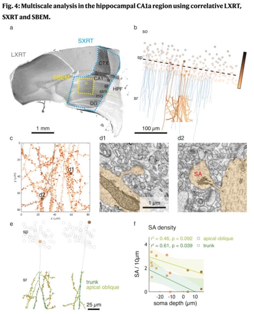
Bosch et al 2022, "Functional and multiscale 3D structural investigation of brain tissue through correlative in vivo physiology, synchrotron microtomography and volume electron microscopy", is an interesting study that brings X-ray microscopy to bear on the problem of correlating structure and function.
The authors studied hippocampal CA1 and olfactory bulb circuits via multiple imaging modalities, including 2-photon calcium imaging, X-ray microscopy, and serial block-face electron microscopy. In all cases, the imaging modalities had different strengths in identifying different circuit elements, and the authors were able to correlate structure and function in interesting ways. The interplay between structural, functional, and molecular-level data will be increasingly critical in systems neuroscience, and this study highlights some important points.
The authors should be commended on showing that X-ray microscopy can be used without causing significant damage on fixed and osmium/uranium/lead en-bloc EM embedded tissue, which is an important advance. The authors also showed that X-ray microscopy can be used at high resolution on thick mammalian brain tissues; this is important because X-ray microscopy has the potential to provide structural details at the level of individual dendrites, which is possible with volume electron microscopy but less easily scalable. Finally, the authors point out that staining protein and lipid distributions defines the ultrastructure of the tissue; this is an important point that is often missed.

