Mouse visual cortex and retinal ganglion cell connectomes
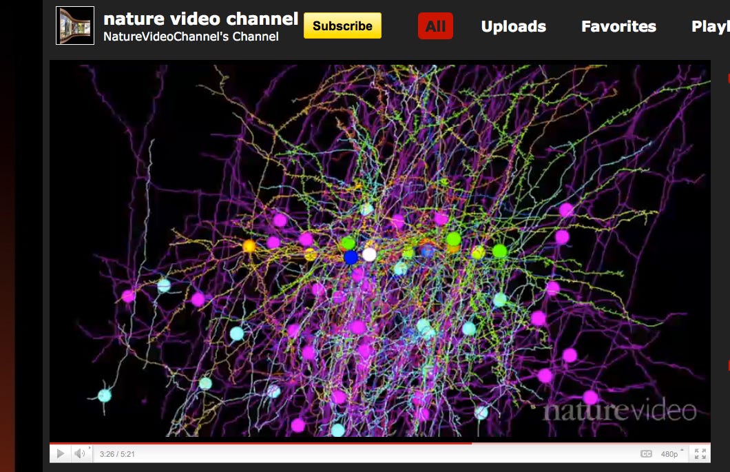
Two papers (here, Bock et al, and here, Briggman et al) published last week, plus a review here by Seung, have helped push the field much further. Click on the screenshot below to watch the 5 min video explaining these studies:

Some say a picture is worth a thousand words, and in that case a 5 min video at ~16 frames/second should be worth ~4.5 million. But I'll still try to use a few words to describe a bit of what these studies accomplished:
The Briggman et al paper studies both the structure and function of one network of neurons. In particular, they look at how direction-selective retinal ganglion cells respond to motion in only one direction. To do this, they first used fluorescence imaging on retinal ganglion cells, while exposing the upstream photoreceptors to moving stimuli. This allows the authors to determine the preferred direction of each of the ganglion cells in their sample.
They then prepped the tissue for electron microscopy and imaged 23 nm (z-direction) slices of a 350 by 300 by 60 um section of it. Then, in what to me seems like the trickiest part of their study, they reconstructed the true synaptic connections between the ganglion cells and their primary inhibitory inputs, the starbust amacrine cells. It turns out that, for a given ganglion cell, almost all of the starburst amacrine cells' input dendrites are oriented opposite to the preferred motion direction, suggesting a clear mechanism for how retinal ganglion cells could maintain their direction selectivity.
The Bock et al paper also focuses on correlating the structure and function of one network of neurons related to vision. In particular, they look at whether the excitatory inputs of inhibitory interneurons in layers 2/3 of the visual cortex come from cells with highly variable preferred stimulus orientations. They did their functional analyses on the stimulus orientation of these cells in vivo, then their perfused the mice and prepped the corresponding volume of the visual cortex for serial section TEM. Their final section had dimensions of 450 by 350 by 52 um and slices of ~45 nm, which contained ~ 1,500 cell bodies.
They located the cells in their EM reconstruction whose stimulus orientation had been determined in vivo. Next, they classified the inputs of the cells on the basis of morphology, and built a "circuit diagram" of the connections. Once they had this diagram, they were indeed able to find many inhibitory interneurons whose excitatory pyramidal inputs had a wide range of preferred stimulus orientations. It's still not entirely clear what these inhibitory interneurons or doing, but this data will help immensely in constructing and falsifying theories about their role.
As Seung discusses in his review, both of these studies are searching for rules of connectivity that can be correlated to the functional properties of those neurons. Ideally, the function of the neurons could be inferred from their connectivity, once we learn more rules of the type that these studies uncover.
He also points out that it would be quite illuminating if we could find an example where a particular type of connectivity allows a neuron to produce a function that is not present in any of its individual inputs. In this way, the long-discussed concept of emergence in neural networks could finally begin to be quantified and partitioned into understandable sub-types.
References
Bock DD, et al. 2011 Network anatomy and in vivo physiology of visual cortical neurons. Nature. doi:10.1038/nature09802
Briggman KL, et al. 2011 Wiring specificity in the direction-selectivity circuit of the retina. Nature. doi:10.1038/nature09818
Seung S. 2011 Towards functional connectomes. Nature doi:10.1038/471170a


