Network motifs in the Drosophila connectome
Cardona et al describe their research towards the Drosophila connectome here. One advantage of studying Drosophila is its lineage tracts, which are groups of neurons in discrete compartments of the brain that differentiated from the same neuroblast early in development. These allow groups of synapses to be broken up into lineage tracts by light microscopy, and analyzed individually. In the following cross section of one hemisphere, one of these lineage tracts is shown in purple:

They aimed for a resolution of 3-4 nm / pixel, which allows them to image synaptic vesicles with diameters of 30-40 nm. These are indicated by arrows in the EM image below:

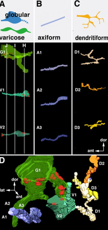
For data analysis, they define a "hierarchy" of objects based on their relationship to one another. This hierarchy ranges from “neuropile compartments" to “neuropile tracts" to “lineages" to “neurons" to "primary branches of a neuron" to "secondary branches of a primary branch." Branches connect to other neurons at synapses, of course. One advantage of the hierarchical object classification is that it will eventually allow the researchers to upload their data sets to neuron modeling programs such as neuron, to simulate the firing of their neurons and try to understand what kind of output the network might produce. Next, they classify the neural projections (dendrites + axons) they image into four types:
axiform ("A," straight, unbranched, 0.2 - 0.4 μm);
varicose ("V," branched, thick are 0.5 - 1.5 μm, thin are 0.15 - 0.4 μm);
globular ("G", variant of varicose with large swellings > 1.5 μm at points, often form end points of axons);
dendritiform ("D," highly branched, often change direction, and < 0.2 μm).
Below are generic models for each, followed by representative 3d digital reconstructions of each type, followed by a 3d digital reconstruction that includes all four types of projections:

Although the same types of projections show up across brain regions, their relative number, direction, and the placement of synapses along them differ between regions. These can form network motifs, which is used in this context to indicate a recurring form, shape, or figure in a design. The most common type of network motif they found they called a "dense overlapping regulatory motif", in which each axon has multiple different targets and each dendrite has multiple different inputs. Less commonly, they found feed forward motifs, in which one projection (neurite) receives input from a second neurite, and both provide output to a common target. Here are schematics of both:

The dense overlapping motif demonstrates how convoluted the Drosophila neural connections can be. In the following example, one presynaptic neurite ("a", bright green) contacts 12 postsynaptic "dendritiform" neurites (yellow-brown) at 3 synapses. Concomitantly, 7 other presynaptic elements (transparent green) contact these same postsynaptic dendritiform neurites. The following shows a representative brain section of the motif as well as front and side views of their reconstructed 3d model:

On the other hand, the feed forward motif underscores how neural signals can be amplified and recombine in a hierarchical manner. In this example, a globular neurite (G) is postsynaptic to a varicose neurite segment (V). Concomitantly, a dendritiform neurite (D, orange) is postsynaptic to both the globular neurite (G) and the varicose neurite (V). Here are two representative brain segments of this motif from the ventral nerve cord cube and a lateral view of the 3d reconstruction:

Not only does this paper have good info about Drosophila neural architecture, but it also shows how researchers are still iterating towards that connectome... Reference: Cardona A, Saalfeld S, Preibisch S, Schmid B, Cheng A, et al. (2010) An Integrated Micro- and Macroarchitectural Analysis of the Drosophila Brain by Computer-Assisted Serial Section Electron Microscopy. PLoS Biol 8(10): e1000502. doi:10.1371/journal.pbio.1000502.


