1: Big study describing the connectome of a 1 mm cubic volume of human brain tissue was formally published. The sample was preserved immediately after it was removed during neurosurgery. It was cut into more than 5000 slices at ∼30 nanometers and imaged with a multibeam scanning electron microscope.
This is the largest connectomics study in human brain tissue to date. 1.4 PB of data. About 57,000 cells and 150 million synapses. Glia outnumber neurons 2:1. Oligodendrocytes are the most common cell type (#LeggoMyOligos).
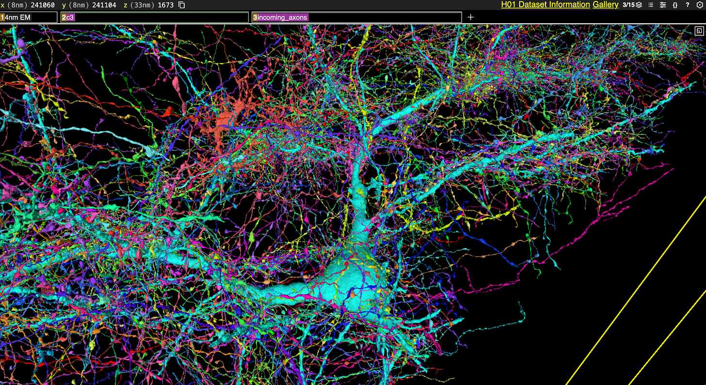
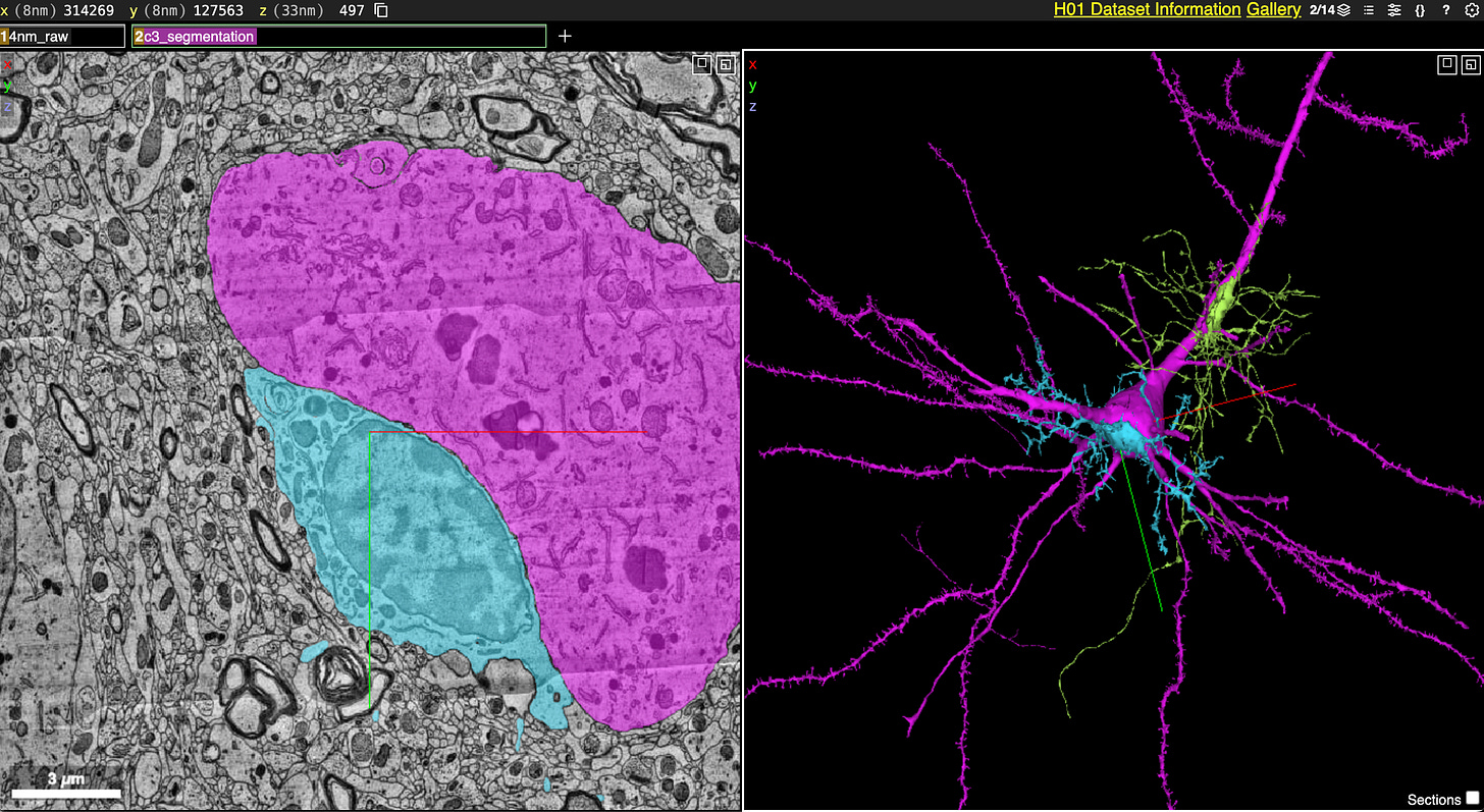
The Neuroglancer visualization data they released is fantastic. Here are all 4000 incoming axons for a pyramidal cell (light cyan):
Here is an example of an “axon whorl”, where an axon (purple) appears to tangle around itself:
Here is an example of a dendrite from one cell tunneling through the soma of another cell, which I didn’t know was possible:
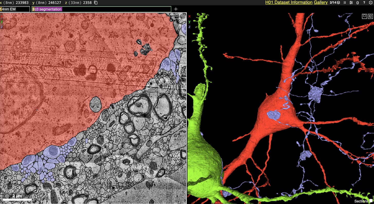
Here they have an example of how many of the glial cells in their data set appear “glued” onto the cell bodies of large pyramidal cells:
This arrangement, with astrocytes processes around neuronal cell bodies, is almost certainly playing a role in how the brain works. It may be related to some kind of glymphatic drainage of the neurons, more explanation here.
Overall, this article is similar to the article published on the pre-print server Biorxiv in 2021. They reported that it took around 4 years to perform the actual work. So this study really reflects the capabilities of the field in 2017-2021.
My understanding is that this team is now working on imaging a 10 mm cubic volume of the mouse brain, as a part of the Brain Connects study.
Why not study more human tissue instead? My guess is that it is too difficult to get such a large volume of brain tissue that is well enough preserved. I realize I’m talking my book, but I think that’s a problem. Studying human tissue is obviously valuable for the study of human disease and this should be prioritized for the upcoming era of connectomics.
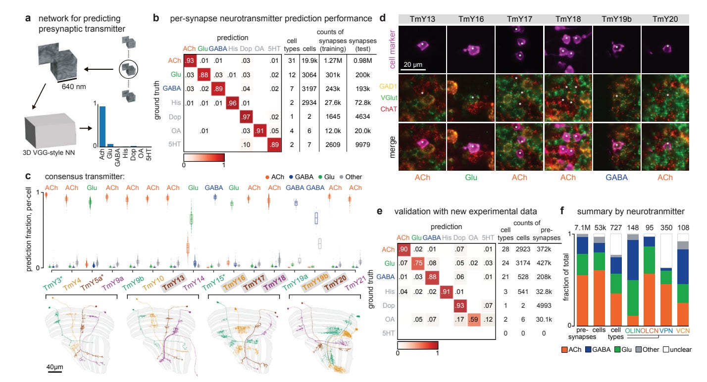
2: Another connectomics study creates an anatomical “inventory” from a FIB-SEM volume of a Drosophila optic lobe, which has ∼53,000 neurons.
One of their many interesting findings is that they are able to predict the presynaptic neurotransmitter based on electron microscopy data with good accuracy:
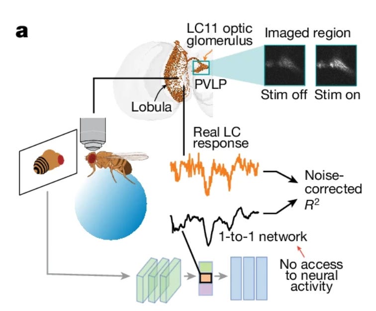
3: A new study uses a cohort of flies with individual neurons genetically silenced to predict neural activity in the fly brain from behavior alone.
The authors recorded the responses of lobula columnar (LC) neurons using calcium imaging in head-fixed male flies viewing visual stimuli. Separately, they also trained a deep neural network model based on behavioral data to predict courtship behavior in these flies. They found a one-to-one mapping between the model units and real neurons.
The model (the “1-to-1 network”) was able to predict the responses of individual neurons to novel stimuli depicting a moving female fly, demonstrating its ability to generalize to unseen visual inputs.
Their model had fairly high predictive accuracy on held out samples, with an R2 of 0.65.
This study reminds me of the idea of “lo fi uploading”, because they aren’t recording any neural activity but they are still able to predict it with some degree of accuracy.
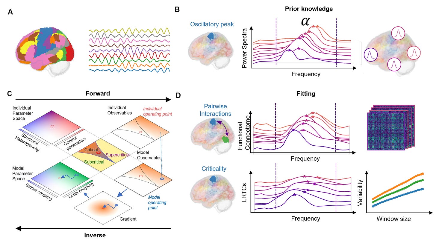
4: New study creates a model that reproduces brain dynamics, including features such as phase synchronization and long-range temporal correlations. This is then fit to magnetoencephalography (MEG) data from individual subjects. This personalized model, which they refer to as a "Digital Twin Brain", can match an individual's phase synchronization profiles to a reasonable degree of accuracy. Early stages but an interesting concept.
5: Study uses a new approach to track the replay of spatial representations in the hippocampus during offline states like sleep. They found that these replayed representations are rapidly reconfigured during the first few hours of post-learning sleep. This reconfiguration predicts how neurons will respond when re-exposed to the same environment in the future.
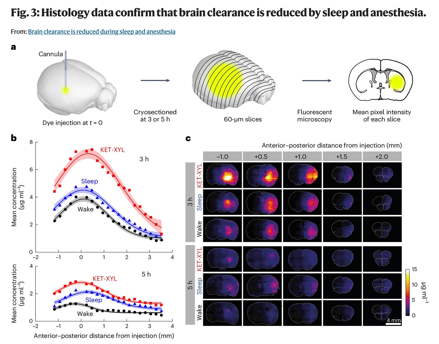
6: New study finds that the clearance of metabolites and toxins is actually reduced in sleep and anesthesia, contradicting previous studies that had reported the opposite. They introduced their dye directly into brain parenchyma, whereas previous studies introduced their dye into the CSF. #MethodsMatter
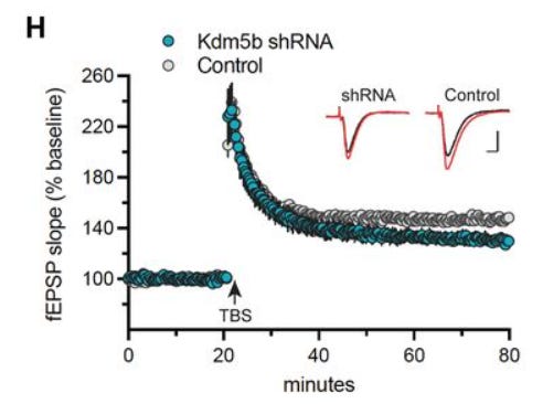
7: Study finds that a risk gene for intellectual disability, Kdm5b, is required for normal memory consolidation and synaptic plasticity in the adult hippocampus. Its knockdown with shRNA leads to deficits in long-term memory and reduces long-term potentiation as measured by field EPSPs in hippocampal slices.
8: New study examines the relationship between genetic risk for depression and 27 adverse life experiences, controlling for ancestry and class background. The depression polygenic index predicts most adversities they tested. The adversities most strongly associated with both the polygenic index and depressive symptoms include disrespectful/insulting events, violent crime victimization, sexual and partner abuse, disability, divorce, incarceration, and unemployment.
It is always difficult to interpret a gene by environment interaction study like this. The authors propose that genetic risk could influence selection into adverse experiences, increasing the risk of depression. It is also possible that depression could affect the way that people recall and perceive negative events in their life.
9: Review finds that only three medications have published evidence that met their quality standards for use in acute anxiety: benzodiazepines, quetiapine (Seroquel) and pregabalin (Lyrica).
10: An immune mechanism for the antidepressant effects of whole body hyperthermia? Reminds me of this profile of Larry David, wherein his daughter noted: “In the house, you’re not allowed to feel bad about yourself or be depressed. He just has no sympathy for it. So if you’re depressed or feeling bad about something, he’ll just tell you to take a shower. That’s like his cure for mental stress. And if it doesn’t work, he’ll be like, ‘Just take another one.’”
I realize I’m a psychiatrist and people sometimes listen to me so I should note that I’m not claiming that the evidence base for hot shower therapy is strong.
11: Advances in stem cell transplants into the brain to treat Parkinson’s disease.
12: Critical perspective on whether we can actually reliably reconstruct images from brain imaging data. Argues that the photo-like reconstructions sometimes presented are actually spurious and due to methodological problems.
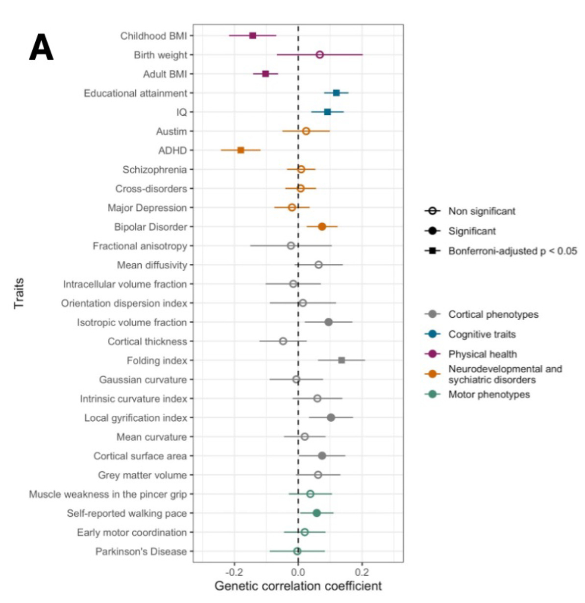
13: GWAS of age of onset of walking. Finds that a later onset of walking has a genetic correlation with higher IQ, higher educational attainment, lower BMI, and lower risk of ADHD.
14: Scott Alexander on what type of theory of mind he would recommend parents teaching their children: "I'm leaning towards "don't". Like yes, they'll absorb the western theory of mind in bits and pieces, but I think this is less bad than deliberately encouraging them to pay attention to their internal landscape."
15: A well-known result in the behavioral psychology literature is the matching law: animals tend to distribute their behavior in proportion to the relative rewards available at each of the different options they are given. But what are the moment-to-moment decision processes that eventually lead to this overall pattern of behavior? A new study shows that trying to figure out these mechanisms is still an active area of research, with a bunch of live theories that may apply in different contexts. Seems potentially relevant to using reinforcement learning to shape the behavior of connectionist machine learning models.
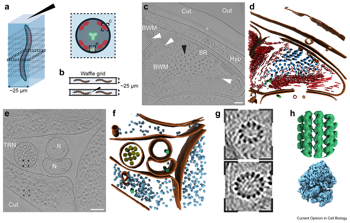
16: Molecular anatomy of C. elegans within cells via cryo-lift-out:
It seems to me that eventually this level of resolution could be eventually reached for mapping across an entire brain. Having access to molecular data would allow for much better inference than just morphological data. Ideally even more detailed molecular data than this, which should also be possible eventually.
17: Cryopreservation of brain tissue has a semi-viral moment this past month after a new method for cryopreserving tissue with four protective chemicals is published. For example, it got thousands of upvotes on this Reddit post, with the title: “Frozen human brain tissue works perfectly when thawed 18 months later | Scientists in China have developed a new chemical concoction that lets brain tissue function again after being frozen.”
Let’s “delve” into their human brain tissue experiment. In it, they found that at least some viable neurons and astrocytes were maintained after the cryopreservation and thawing process, allowing for outgrowth and tissue culture. This is a nice result. However, I'm not sure how this is different from previous studies that have shown that human brain tissue can be cryopreserved, rewarmed, and allow for viable cells to grow (for example, here or here).
It’s always interesting seeing which findings catch the attention of the public and which do not. Regardless, I am heartened to see more research and interest in this area.
18: New brain archiving center at the University Hospital Erlangen, led by Dr. med. Alexander German. They report that they are performing MRI and electron microscopy on the samples and also preserving them for the long-term. Here is an English translation of the text on their website:
“What are the consequences of brain archiving?
If you make your brain donation available for research, you help to improve the prevention, detection, and treatment of diseases. With new technical possibilities, mapping the brain structure (connectome) could become possible in the future. The consequences of this are currently not foreseeable. It is speculated that after brain archiving, it could become possible in the distant future to read out memories collected during one's lifetime or to regain brain function. The aim of the brain archive is to be open to this perspective on brain specimens as well. You decide on the uses for your brain specimen. At present and for the foreseeable future, however, no functional preservation or restoration of brain function will be possible. Currently, the research purposes for brain specimens are not fully known. There could be a significant increase in the demand for brain specimens, e.g., in the future when studying biological intelligence and for the safety of advanced artificial intelligence (AI). Permanent storage is ensured by CeBE [Central Biobank Erlangen]. You can optionally limit the storage period.
Who can participate in the brain archive?
Adults capable of giving consent can participate in the brain archive, regardless of whether they are healthy or have a serious illness, for example. There are no changes whatsoever during one's lifetime as a result of consenting. Brain archiving raises philosophical and also spiritual questions. Therefore, the establishment of the brain archive is carried out in cooperation with the Faculty of Humanities and Social Sciences at FAU [Friedrich-Alexander-Universität], in order to accompany the diverse conceivable questions of brain archiving and to take into account the differences between various age groups, religious beliefs, and worldviews.
Important: A declaration of consent to participate can be revoked at any time without giving reasons.”
Congratulations and Godspeed to all involved in this historic effort.


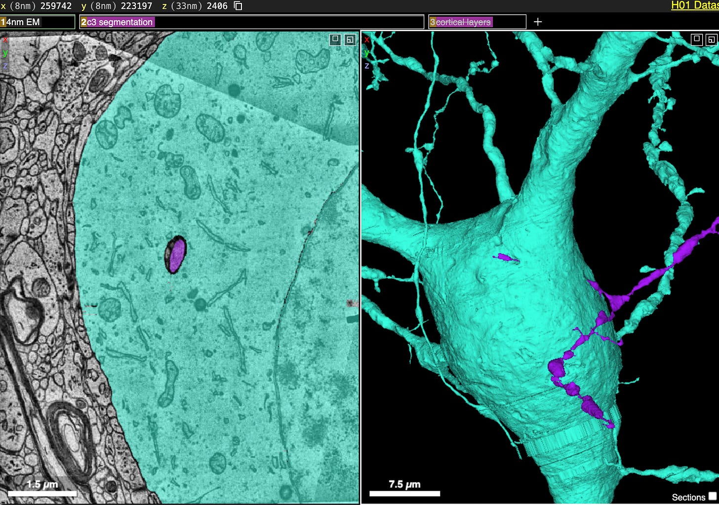

Regarding #1: I spoke to one of the authors last week, and he thinks that the "dendrite from one cell tunneling through the soma of another cell" is probably an image segmentation or tracing bug, rather than a real phenomenon.
Wow - beautiful data.